

Infrastructures
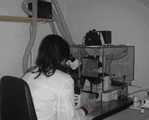
Advanced Light Microscopy
Equipment:
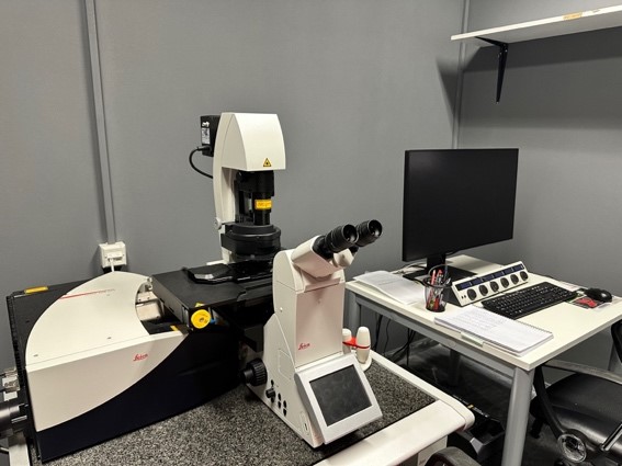
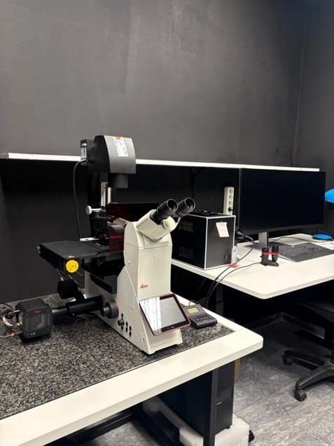
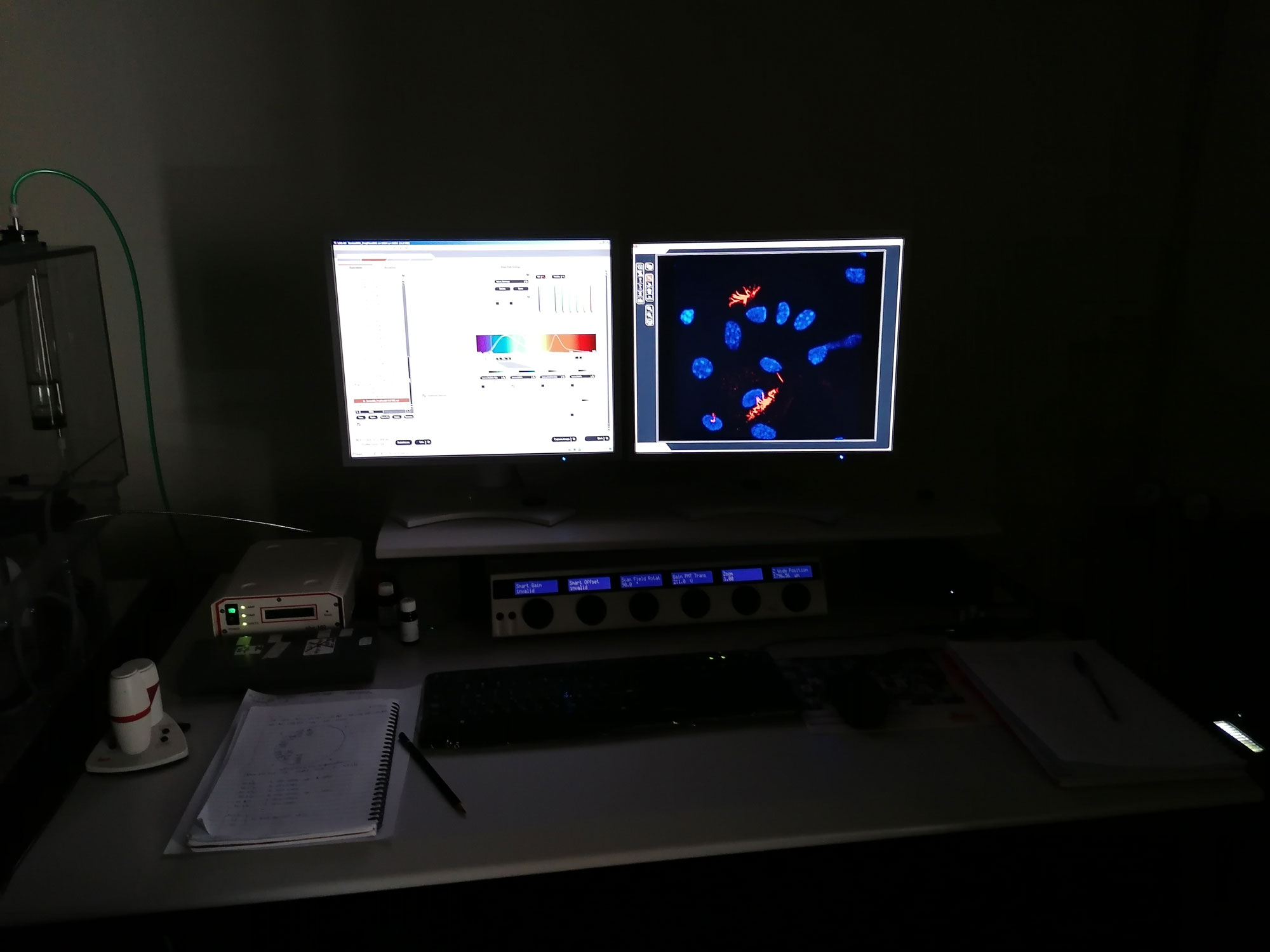
Leica TCS SP5 Confocal Microscope equipped for live-cell imaging
Leica TCS SP8 Multiphoton Microscope
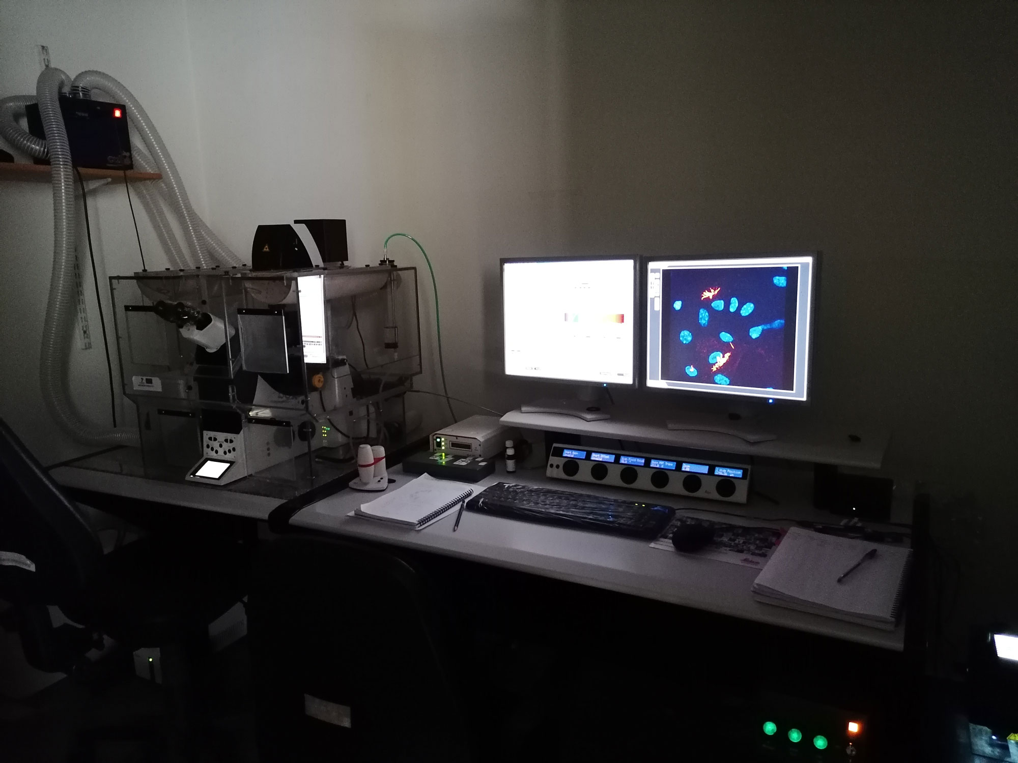
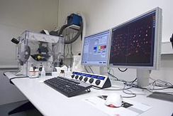
Leica DMI8 “Thunder Imager”
Supported Methodologies:
Confocal Microscopy, Epifluorescence Microscopy, 3D reconstruction, Time-lapse Microscopy, Fluorescence Recovery After Photobleaching (FRAP), Fluorescence Loss in Photobleaching (FLIP), Förster Resonance Energy Transfer (FRET), localized DNA damage, super-resolution processing
Applications:
Co-localization of multiple fluorophores within cells and tissues, functional imaging, proteininteractions in living cells, stem cell analysis, cell-based assays for drug validation.
Animal House
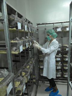
The Animal House in the University of Patras is a fully equipped facility located in the ground floor of the Medical School where animals are kept in conditions with regulated ventilation, temperature, light and relevant humidity, monitored on a 24 hour basis. There are housed more than 3500 small animals including transgenic and knockout mice, rats and rabbits used, for research purposes by members of the Patras Medical School. The Animal House includes a fully equipped surgical room, with facilities for in utero electroporation and stereotactic injection, as well as rooms equipped for metabolic experiments. High standards of biosafety are preserved in all rooms, while the use of animals follows the 3R guiding principles (Replacement, Reduction and Refinement).
In Utero Electroporation
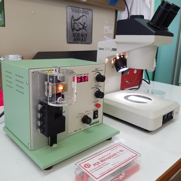
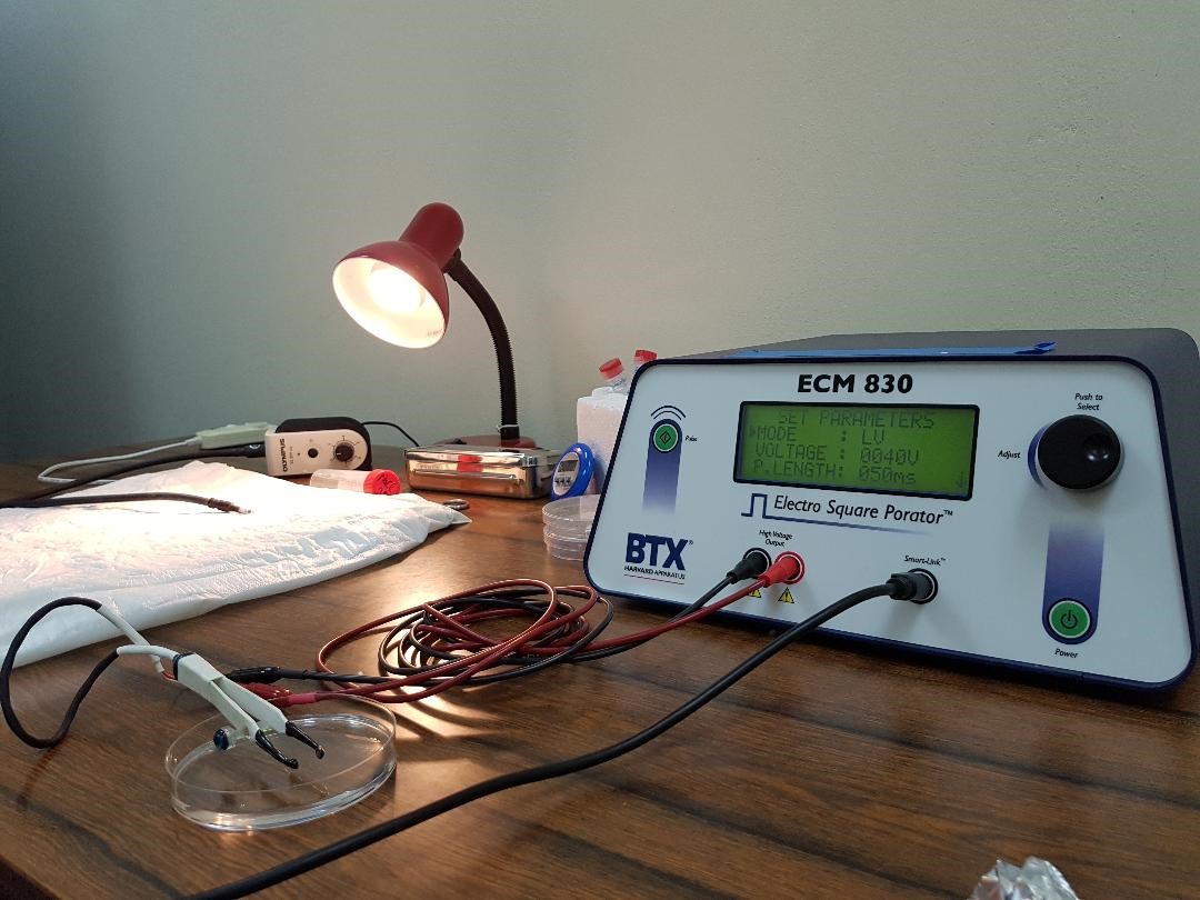
In utero electroporation technique has been established a few years ago from our Research Team in the Animal House at the University of Patras. In our laboratory we take advantage of the technique in order to study brain development and neurogenesis. Between lines, we introduce plasmid DNA into the brain ventricles of embryos from a timed female pregnant mouse, aiming to overexpress or delete a specific gene, whose function we wish to study. More specifically, DNA of interest is introduced into the mouse brain, followed by the application of an electrical pulse in order to guide DNA to a specific brain region. Therefore, we are able to study genes function in vivo without the need of a specific transgenic mouse line. The device used for the electroporation is the Electron Square PoratorTM ECM830 by BTX® HARVARD APPARATUS. The 1-10 μl micropipettes, are prepared with Narishige PC-10 micropipette puller, a dual-stage micropipette processing system which provides reliable and consistent production of micropipettes by pulling the glass capillary vertically.
Microarray Analysis
Εquipment:
Microarray Scanner Scan Array Express(PerkinElmer), Hybridiser HybArray12 (PerkinElmer), Bioanalyser 2100 (Agilent)
Supported Methodologies:
Microarray Analysis, Microanalysis
Applications:
High-throughput gene expression analysis, Genotyping, SNP Analysis
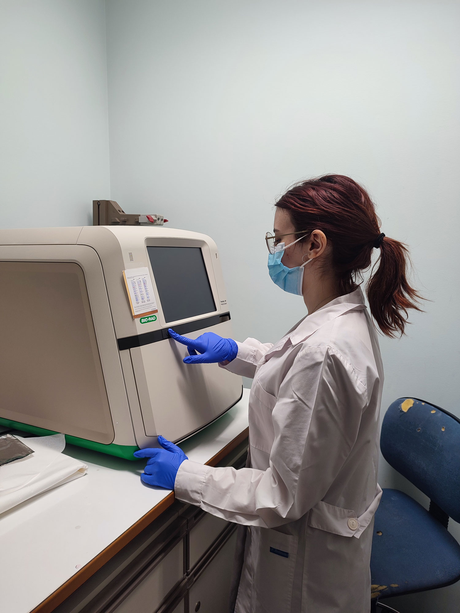
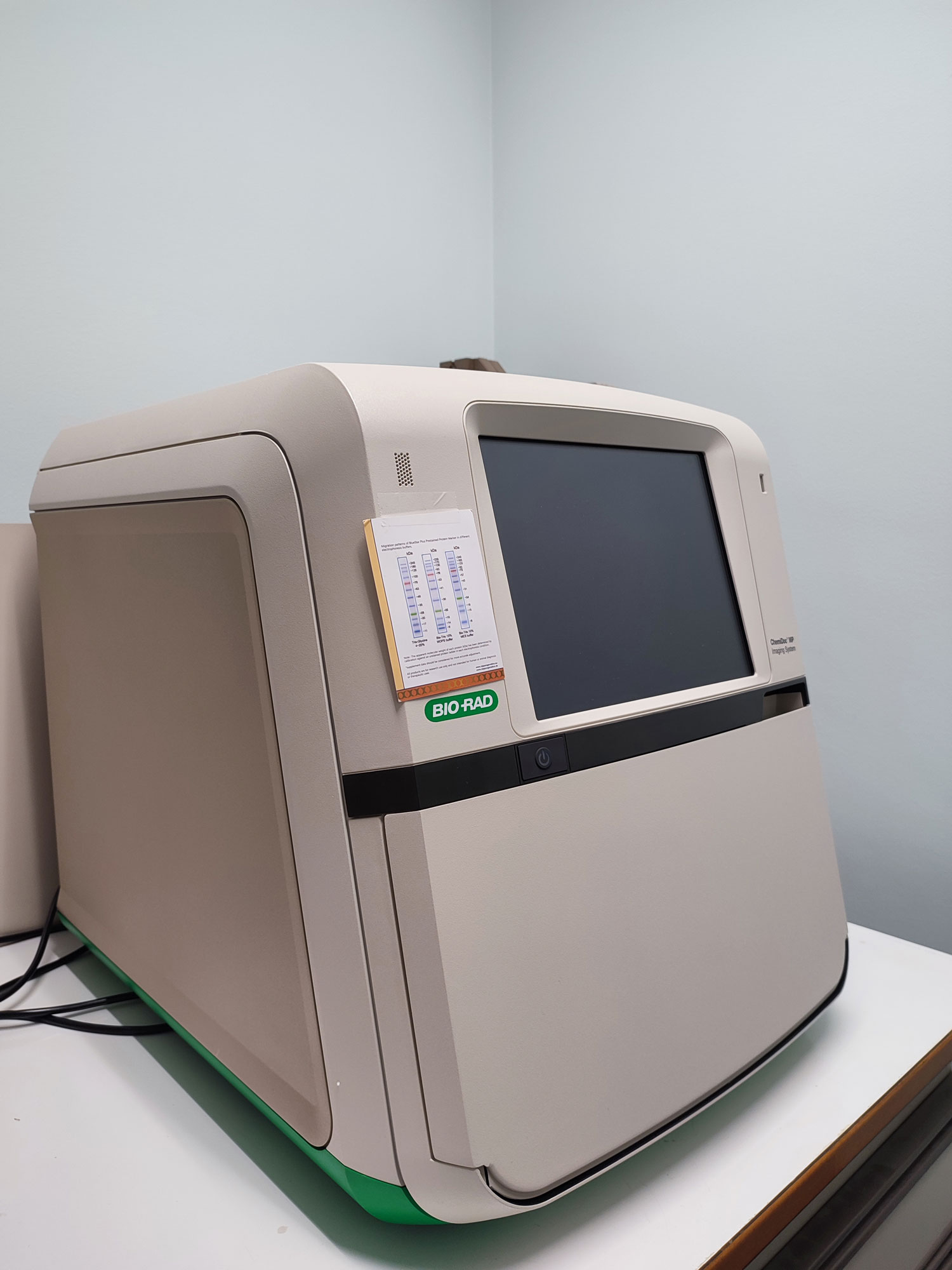
Modern Blot and Gel Imaging System
Εquipment:
Chemidoc MP Imaging system, Biorad
Applications:
Western Blot, 2-D Electrophoresis, Imaging and Analysis, Horizontal Nucleic Acid Electrophoresis, Stain-Free Imaging Technology
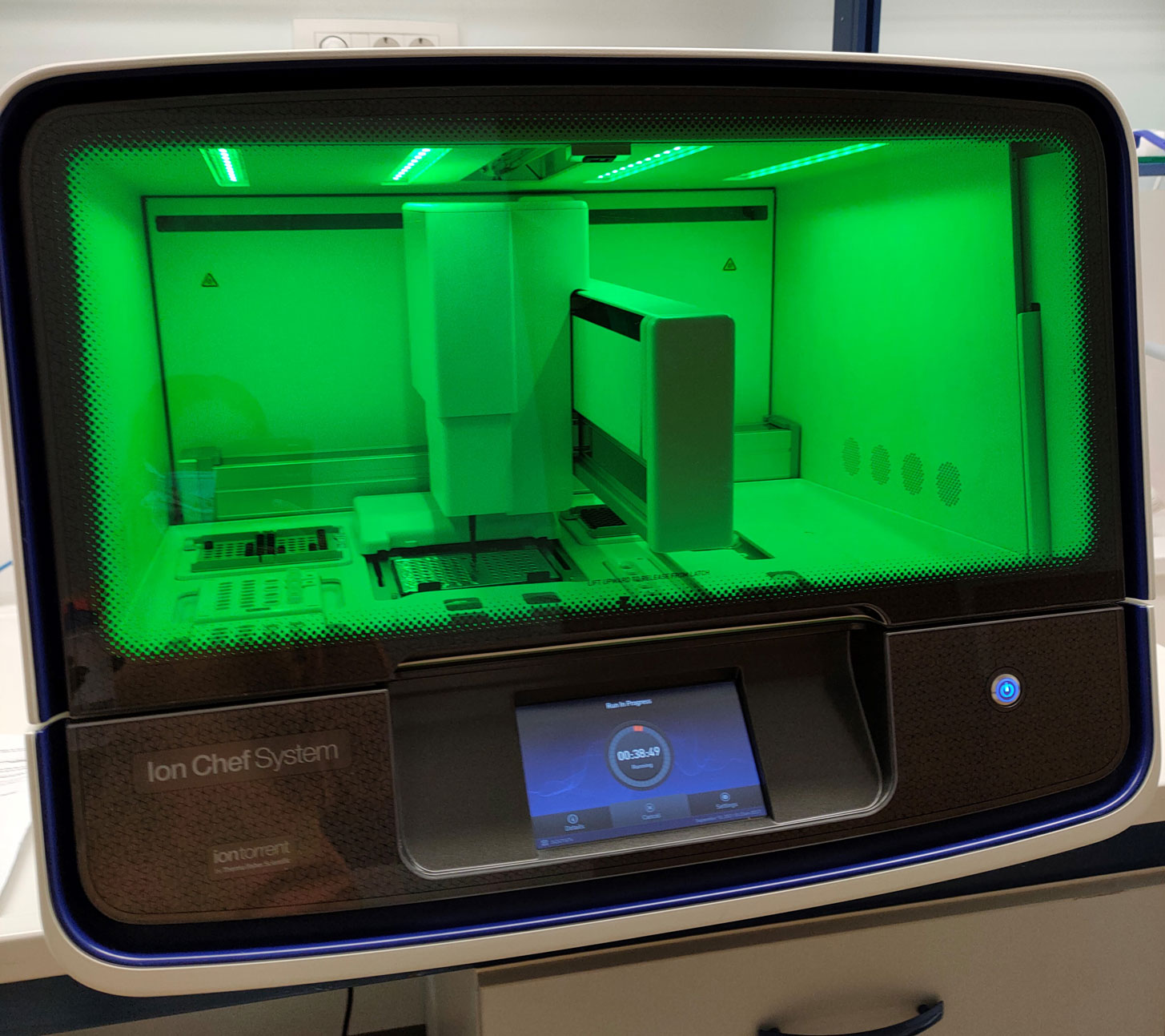
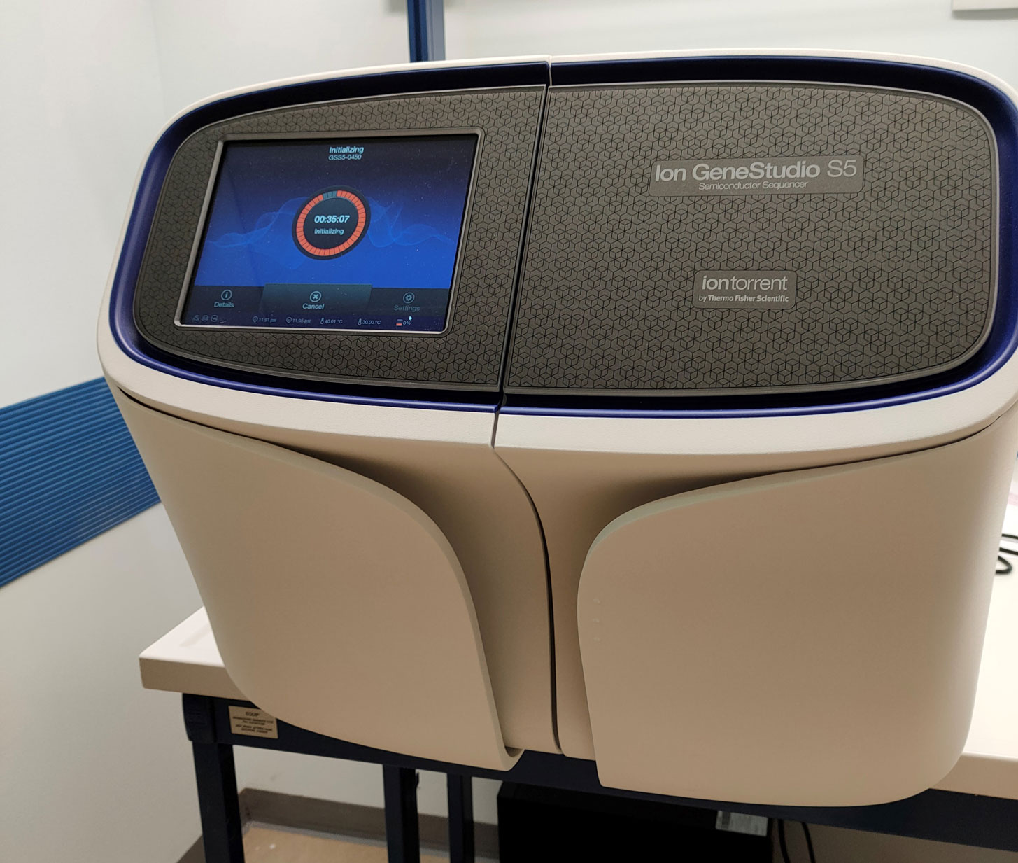
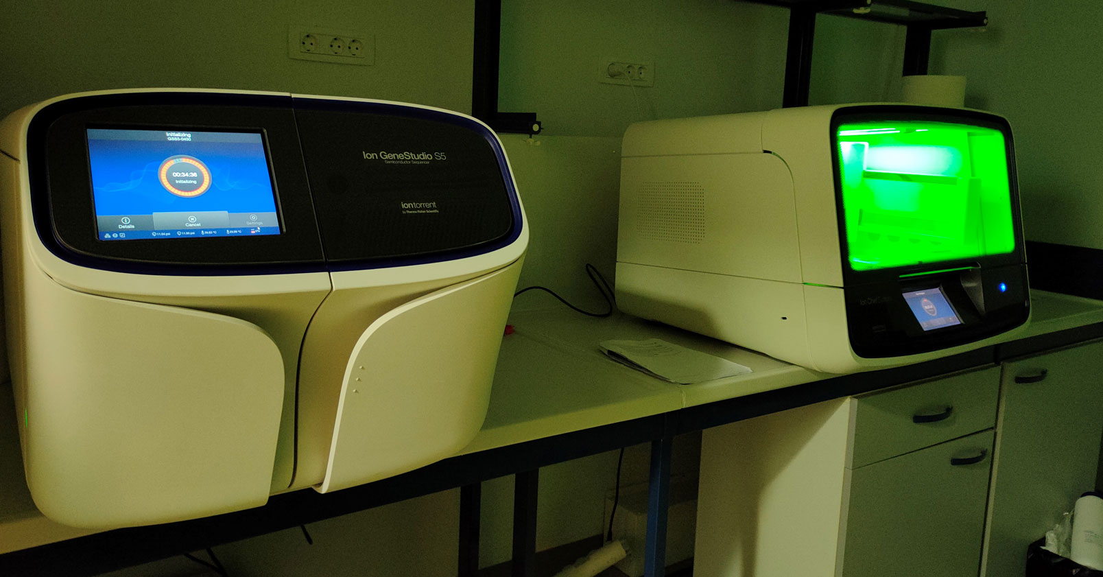
DNA Sequencing System
Εquipment:
Ion GeneStudio™ S5 System, Thermo Fisher Scientific
Applications:
Ion Torrent™ Next-Generation Sequencing, SARS-CoV-2 Pathogen Research Solutions
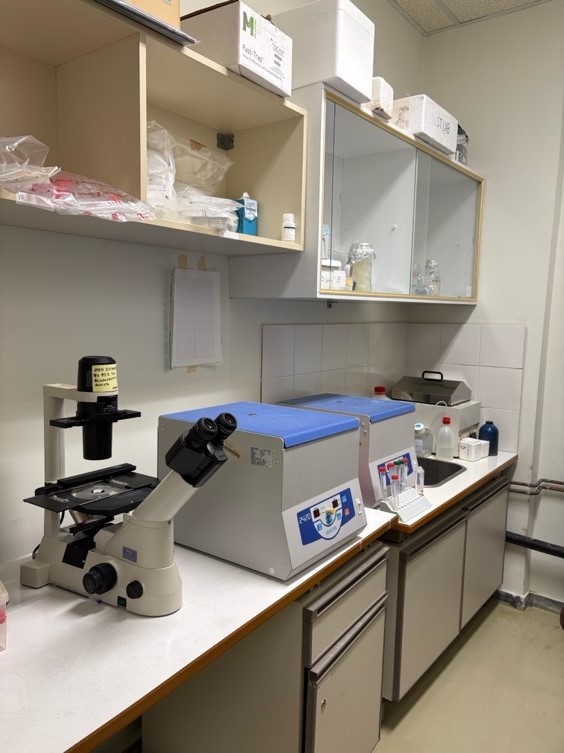
Tissue Sectioning
Εquipment:
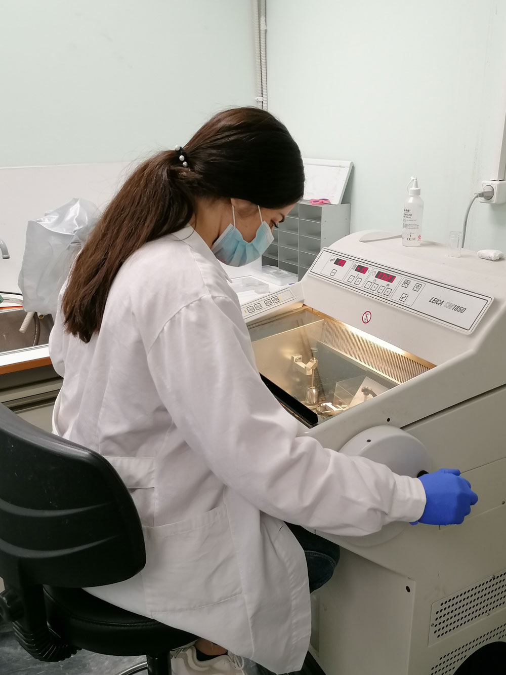
Leica CM1850 Cryostat
The cryostat is a specialized instrument composed of a tissue microtome (cryotome) installed within a cooling chamber that allows for rapid freezing of tissues and a continuous process of cutting tissue at low temperatures (typically around − 15 to −30°C), which can later be used in Immunofluorescence assays.
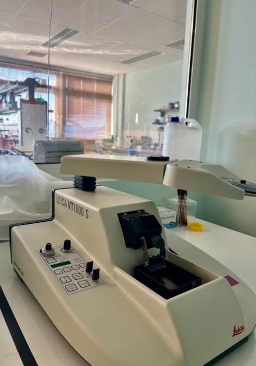
Leica VT1000S Vibratome
The vibratome uses a rapidly vibrating blade to slice fresh (or fixed) biological tissues (like brain, kidney, liver) into thin, uniform sections (20-1000 microns thick) while keeping cells alive and structures intact
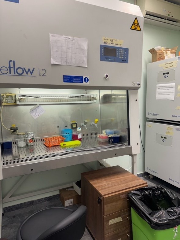
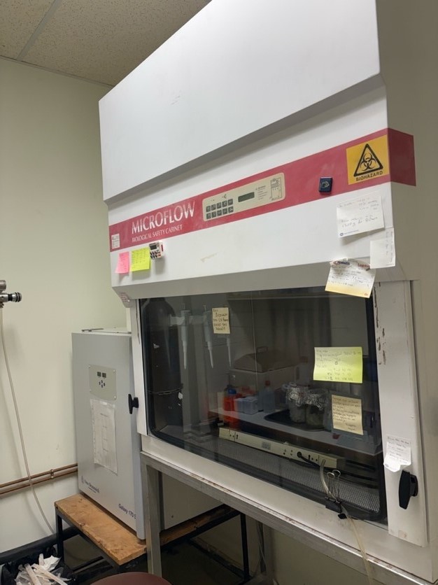
Tissue culture
Biosafety level 1 tissue culture
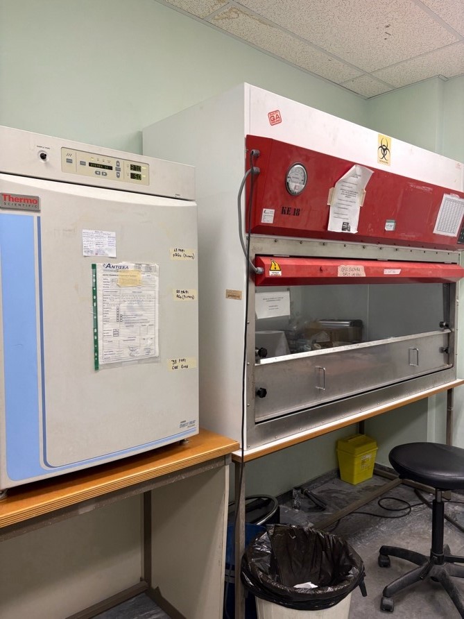
Biosafety level 2 tissue culture – Lentiviral culture
Flow Cytometry and Cell Sorting
Εquipment:

FACS Aria III – cell sorter
The FACSAria III is a high-performance, multi-laser, digital cell sorter for rapid, precise analysis and isolation of specific cell populations, known for its multicolor capabilities, high speed (up to 70,000 events/sec), and flexibility with two lasers and multiple nozzle sizes for various cell types, enabling complex sorting into plates or tubes with excellent sensitivity and purity. It’s a powerful tool for isolating rare cells, supporting extensive fluorescent parameter detection for advanced research.
FACS Celesta – cell analyzer
The BD FACSCelesta is a compact, benchtop flow cytometer designed for easy, multicolor cell analysis, especially optimized for bright BD Horizon Brilliant dyes, using multiple lasers (violet, blue, red) to detect many fluorescent parameters for deep insights into cell populations.