1. The pup is sacrificed by cutting the head off instantaneously, the brain is extracted and placed into in a 30-mm Petri dish with ice-cold dissection medium (10mM of HEPES in HBSS 1×).
2. The brain is divided and a coronally-oriented cut is made at the posterior-most aspect of the two hemispheres. The ventricle from the caudal side is opened up using forceps, allowing to visualize the ventricular face of the wall of the lateral ventricle.
3. Subsequently, the lateral wall is completely exposed and sub-dissected using fine forceps.
4. The lateral walls from one pup are mechanically dissociated in dissociation medium and centrifuged in 1100rpm for 4min (RT).
5. The supernatant is carefully removed and the pellet containing the cells is re-suspended in pre-warmed (37 °C) culture medium (DMEM-High Glucose 4.5 GlutaMAX, 10% FBS and 1% Pen/Strep).
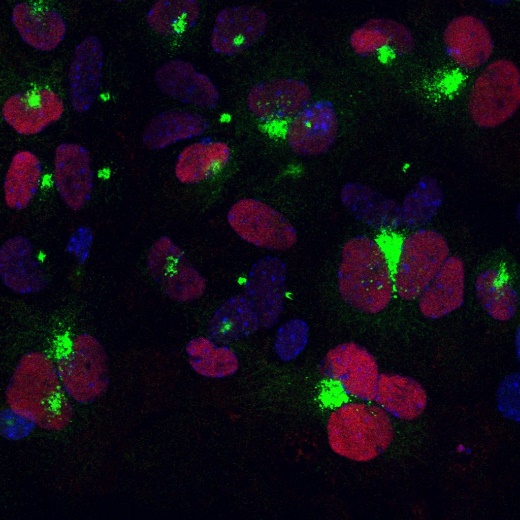
6. The cells are seeded onto poly-D-lysine–coated glass coverslips. Note: The yield from one lateral wall is seeded on each well.
7. When cells reach 90%–100% confluence (3 days after plating), media were switched to 2% FBS (other ingredients same as above) and not changed again for the duration of experiment.
Protocol modified from Paez-Gonzalez et al., Neuron, 2011
This work was supported by Aristeia II, GEMCCTR “Self-renewal and differentiation decisions in neural stem cells: Geminin, cell cycle control and transcriptional regulation”